
Kidney structure and function
|
The kidneys are organs of the urinary system which are fundamental in controlling the water content of the body by removing excess water, salts and the waste product urea.
Blood circulates the body under pressure and is brought to the kidney in the renal artery. The kidneys filter the blood and then reabsorb all the useful materials such as glucose. After it has been purified the blood returns to the circulation through the renal vein. The kidney makes urine from the excess water, salts and urea. Urea is a waste product produced in the liver when surplus amino acids are broken down called deamination. Urea becomes toxic at high concentrations and must be removed from the blood. Urine travels away from the kidneys in tubes called ureters. The urine is stored in the bladder. When the bladder is full the dog gets the urge to expel the contents through the urethra. |
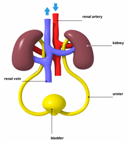
Simple diagram of the Urinary system
|
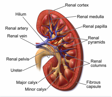
Diagram of human kidney - Human kidneys are multipapillary. Canine kidneys are unipapillary and have a single crest-like papilla produced by the fusion of the renal lobes
|
|
|
Urine is produced in microscopic tubes in the kidney that are called the nephrons that are found in the medulla.
Normal kidneys have many millions of nephrons. Chronic kidney failure results in the progressive loss of these structures together with the production of fibrous tissue and cysts within the kidney tissues. The process of urine production begins in the nephron when the glomerulus filters the blood under high pressure into the Bowman's capsule - almost all the contents of the blood except the blood cells and large protein molecules are pushed out of the blood forming a glomerular filtrate. This liquid contains large amounts of water, glucose, salts and urea. Large molecules such as protein are usually too large to fit through the blood capillary walls. If there is a problem with the structure of the kidney then large protein molecules can be make their way into the filtrate. The bowman's capsule collects the filtered liquid and it enters the tubules. In a normally functioning kidney all the filtered glucose is reabsorbed immediately back into the blood capillaries. As the rest of the filtrate travels through the tubules water and salts that are still needed by the body are reabsorbed into the blood thereby concentrating the urine. If the kidney is not functioning correctly the urine will not concentrate as the water is not reabsorbed. This produces a dilute urine with a low Urine Specific Gravity (USG). The remaining waste that contains the excess water, excess salts and urea, is now called urine. |
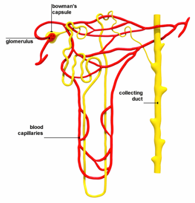
Structure of a Nephron
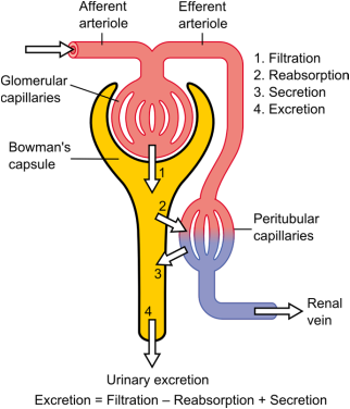
Diagram showing the function of the nephron
|
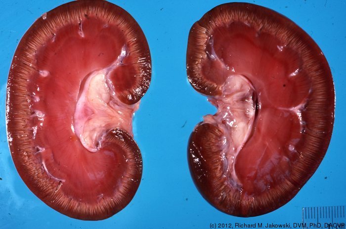
Normal kidneys
|
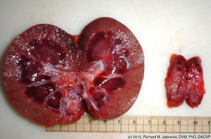
Unilateral Dyplasia of a dog showing the difference in a severely affected right kidney and the left
|